Figure 2.

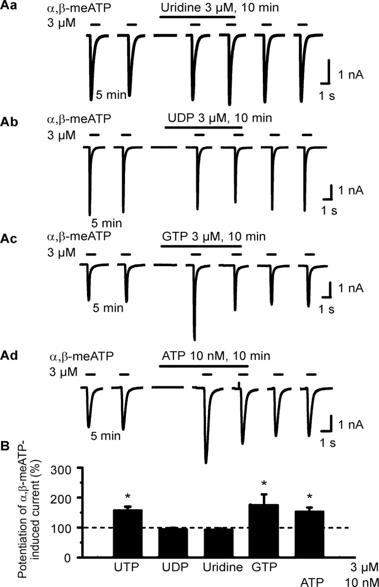
Effects of extracellular nucleotides and nucleosides on α,β-meATP-induced currents in HEK293-hP2X3 cells. The experimental procedures were similar to those described for Figure 1 B. Patch pipettes filled with GDP-β-S (300 μm)-containing solution were used. Superfusion with agonists started 5 min before the third application of α,β-meATP. A, Original tracings show that the α,β-meATP currents were potentiated by GTP (3 μm; Ac) and ATP (10 nm; Ad) but not uridine (Aa) or UDP (3 μm each; Ab). B, Mean ± SEM of 4-11 experiments similar to those shown in A.*p < 0.05, statistically significant difference from 100%.
