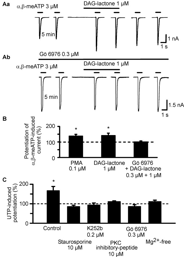
Figure 3.
Effects of PKC activators on α,β-meATP-induced currents in HEK293-hP2X3 cells; interaction of PKC inhibitors with PKC activators and UTP. The experimental procedures were similar to those described for Figure 1 B. Patch pipettes filled with GDP-β-S (300 μm) containing solution were used. Superfusion with PKC activators (PMA and DAG-lactone) started 5 min before the third application of α,β-meATP. Superfusion with PKC inhibitors (Gö 6976, staurosporine, K252b, and PKC inhibitor peptide) and procedures known to block the PKC-mediated phosphorylation reaction (Mg2+-free external medium) started 10 min before the first application of α,β-meATP and continued for the duration of the whole experiment. A, Original tracings show that the α,β-meATP (3 μm) currents were potentiated by DAG-lactone (1 μm;Aa) in the absence of Gö 6976 (0.3 μm) but not in its presence (Ab). B, Potentiating effect of PMA (0.1 μm) and DAG-lactone alone, but not DAG-lactone in combination with Gö 6976, on the α,β-meATP current. Mean ± SEM of 7-12 experiments similar to those shown in A. C, Blockade of the potentiating effect of UTP (3 μm) on the α,β-meATP (3 μm) current by staurosporine (0.1 μm), K252b (0.2 μm), PKC inhibitor peptide (10 μm), Gö 6976 (0.3 μm), and a nominally Mg2+-free external medium. Mean ± SEM of 6-10 experiments similar to those shown in A. *p < 0.05, statistically significant difference from 100% both in B and C.

