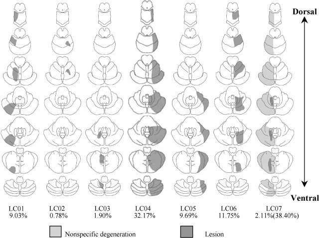
Figure 1.
Cerebellar lesions in the seven patients. MRI scans were analyzed by a neurologist, and the extent of pathology was sketched on seven axial cerebellar slices. Four lesions resulted from damage associated with stroke (LC01, LC02, LC04, LC06), and three lesions resulted from damage associated with tumor resection (LC03, LC05, LC07). Below each template is the percentage of cerebellum lesioned. In patient LC07, the first value relates to lesion damage, and the second value relates to nonspecific degeneration.

