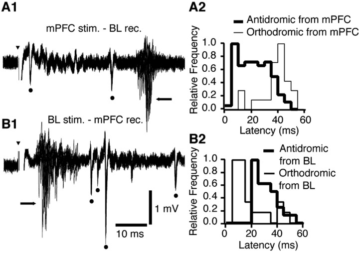
Figure 4.
The axons of BL neurons projecting to the mPFC have a shorter conduction time than mPFC axons ending in the BL nucleus. A1, Example of multiunit responses elicited by mPFC stimuli in the BL nucleus (20 superimposed sweeps). A2, Distribution of antidromic (thick lines) and orthodromic (thin lines) response latencies (all experiments combined). Each distribution was normalized to its mode. Antidromic response latencies ranged widely from 4 to 46 ms, with a mode of 8 ms. B1, Example of responses elicited by BL stimuli in the mPFC (20 superimposed sweeps). B2, Distribution of antidromic (thick lines) and orthodromic (thin lines) response latencies (all experiments combined). Antidromic response latencies ranged from 21 to 47 ms, with a mode of 24 ms. Dots and arrows mark antidromic and orthodromic responses, respectively. Bins of 5 ms in A2 and B2. Arrowheads indicate stimulation artifacts.

