
Figure 6.
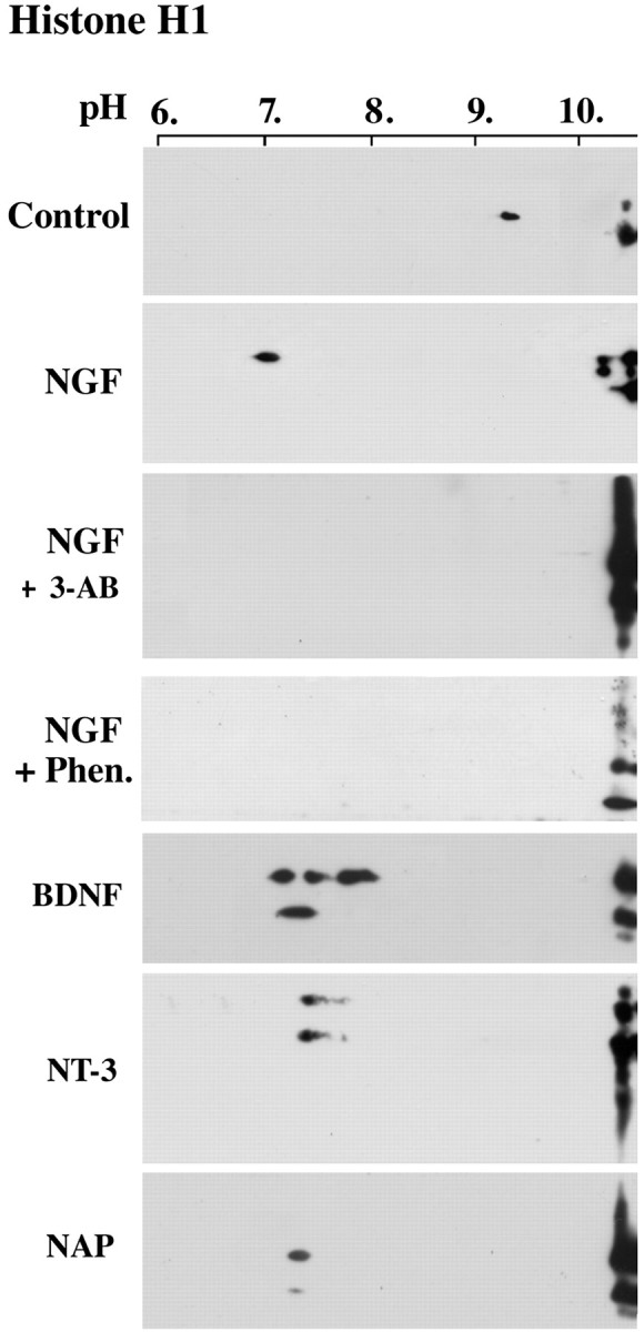
PolyADP-ribosylation of linker histone H1 in neurons treated by nerve growth factors and NAP. PolyADP-ribosylation of histone H1 in nuclei isolated from cortical neurons treated by neurotrophins, as indicated. PolyADP-ribosylation was assayed by the shift in the pI of H1 toward acidic pH values. The shift in pI was prevented by the PARP-1 inhibitors 3-AB (0.5 mm) and Phen. (25 μm). Brain cortical neurons were incubated for 5 min with 100 ng/ml NGF, BDNF, NT-3, or 10-12 m NAP (n=3 for each treatment). Histone H1 was labeled by monoclonal anti-human H1 antibody (Upstate Biotechnology, Lake Placid, NY) on Western blots of nuclear proteins separated by two-dimensional gel electrophoresis (see Materials and Methods).