
Figure 7.
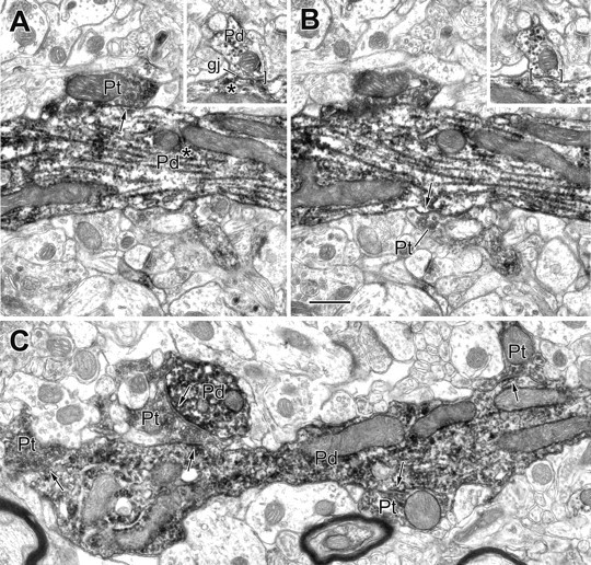
Electron micrographs of PV+ dendrites exhibiting a gap junction and receiving synapses from PV+ terminals. A, B, Two serial sections of a large-caliber PV+ dendrite (Pd) showing two of three PV+ terminals (Pt; arrows) that make synaptic contact with this dendrite. The inset depicts a gap junction (gj) involving this dendrite and a small PV+ dendrite (Pd; brackets) that was located 11 μm from the synapses. The asterisks indicate that all contacts are with the same PV+ dendritic trunk. C, Longitudinal view of a varicose PV+ dendrite (Pd) receiving synaptic input from four PV+ terminals (Pt; arrows). One of these PV+ terminals also synapses onto an adjacent PV+ dendritic cross section (Pd; arrow; top left). Scale bar, 0.5 μm.