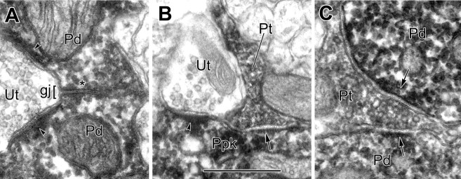
Figure 8.
Comparison of the morphology of an IP-labeled gap junction with IP-labeled chemical synapses. A, Detail of the IP-labeled PV+ dendrites (Pd) shown in Figure 2C in gap junctional contact (gj; asterisk). Each dendrite receives an asymmetrical synapse (arrowheads) from an unlabeled terminal (Ut). B, Detail from Figure 6 A of an IP-labeled PV+ perikaryon (Ppk) receiving synaptic contacts from a PV+ axon terminal (Pt; arrow) and unlabeled axon terminal (Ut; arrowhead). C, Detail from Figure 7C of an IP-labeled PV+ terminal (Pt) making synaptic contacts (arrows) with two PV+ dendrites (Pd). Note that peroxidase reaction product fills the central gap of the gap junction, whereas an unstained synaptic cleft separates the outer leaflets of presynaptic and postsynaptic plasma membranes of chemical synapses. Scale bar: (in B) A-C, 0.5 μm.

