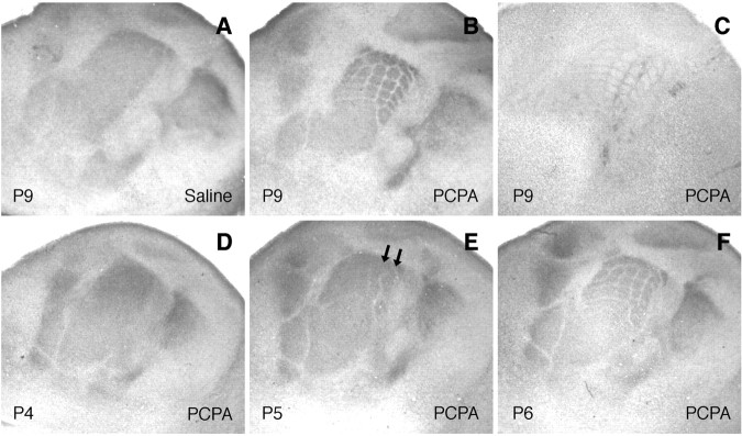
Figure 1.
Rescue of large barrels in MAOA-KO mice treated with PCPA. Tangential sections of flattened hemispheres of MAOA-KO mice are shown. Mice were treated with saline (A) or PCPA (B-F) treatment from P2. 5-HTT immunohistochemistry (A, B, D-F) reveals the sensory thalamocortical fibers and Nissl staining (C) in the layer IV neurons. A-C, A rescue of barrel clustering in the large whisker representation is noted at P9 in PCPA-treated mice (from P2 to P8; B, C) but not in the saline-treated mice (A). D-F, In MAOA-KO mice treated from P2 on, no segregation of fibers is visible at P4 (D), rows A and B (arrows) corresponding to the large whiskers are visible at P5 (E), and individual barrels are observed at P6 (F).

