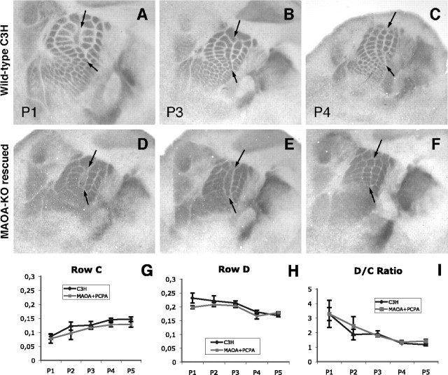
Figure 3.
Critical period for lesion-induced plasticity is unchanged in MAOA-KO rescued mice. Quantification of the row C lesion effect is shown. A-F, Row C whiskers were electrocauterized at P1 (A, D), P2, P3 (B, E), P4 (C, F), and P5 in C3H mice (A-C; n = 40) and MAOA-KO rescued mice (D-F; n = 34). A, D, Lesions at P1 cause a decrease of row C and an expansion of adjacent barrel rows B and D cortical areas in both C3H (A) and MAOA-KO rescued (D) mice. B, E, The lesion still has a milder effect at P3. C, F, No effect is visible when row C lesion is made at P4 and assessed with 5-HTT immunolabeling. G-I, Quantification of lesion effect. The areas of rows C (G) and D (H) were compared with the total area of the large barrels for normalization. A, At P1, the row C area is low and increases progressively with age until P4 in MAOA-KO rescued and normal mice. B, Conversely, the row D area decreases with age from P1 to P4 in wild-type mice. A slight difference can be observed because the row D area seems grossly identical from P1 to P3 in MAOA-KO rescued mice. A decrease is clearly observed between P1 and P4, as well as stabilization in relative area size from P4 to P5 in both genotypes. The D/C ratio plasticity index clearly indicates a general decrease from P1 to P4 and stabilization thereafter, stating the absence of plasticity after P3. C3H mice values are in black, and MAOA-KO rescued mice values are in gray. Arrows indicate row C. Error bars represent SEM.

