
Figure 1.
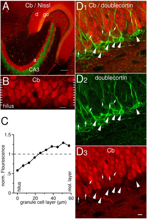
Distribution of calbindin-D28k immunoreactivity in the dentate area. A, Dual channel scan obtained with a confocal laser microscope. Calbindin-D28k (Cb) is present throughout the entire cytoplasm of granule cells: dendrites (d), somata (gc), and axons (a) are stained (red). Note that CA3 pyramidal cells are completely negative, whereas some scattered interneurons are found positive. Green indicates fluorescent counterstain (“Nissl”). Scale bar, 100 μm. 10× objective. B, High-magnification scan of the granule cell layer stained for calbindin-D28k. Note that the calbindin-D28k content of granule cells is heterogeneous: granule cells adjacent to the molecular layer (top) are brighter than hilar dentate granule cells (bottom). The white ticks on the sides of the image indicate how the scan was divided into 10 sections for quantitative analysis in C (see Materials and Methods for details). C, Quantitative analysis of the gradient of calbindin-D28k immunoreactivity across the granule cell layer. The granule cell layer (n = 10) was divided into 10 sections, and the normalized mean fluorescence in each section is plotted versus its position within the layer. D, Double-labeling with anti-calbindin-D28k (red) and anti-doublecortin antibodies (green). Cells that are weakly calbindin-D28k-labeled are doublecortin-positive (small arrows). Strongly calbindin-D28k-positive cells are doublecortin-negative. Note that cells intensely stained for doublecortin appear calbindin-D28k-negative (large arrowheads).