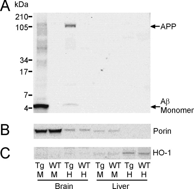
Figure 5.
SDS-PAGE and Western blot analysis of APP and Aβ in brain mitochondria of Tg2576 mice. A, Aβ and APP in total homogenate (H) and mitochondria (M) samples from the brain and liver of 18-month-old Tg2576 (Tg) and wild-type (WT) mice were resolved on 10-20% Tris-tricine gels and Western blotted with WO2 for Aβ/APP. Aβ was greatly enriched in the brain mitochondria of Tg2576 mice but nondetectable in brain mitochondria of age-matched WT controls. Fifty micrograms of total protein (for mitochondria samples) or 30 μg of total protein (for homogenate samples) was loaded per lane. The gels were reprobed with Porin for mitochondria (B) or HO-1 for endoplasmic reticulum (C), and this revealed that the mitochondria fractions were relatively free from endoplasmic reticulum.

