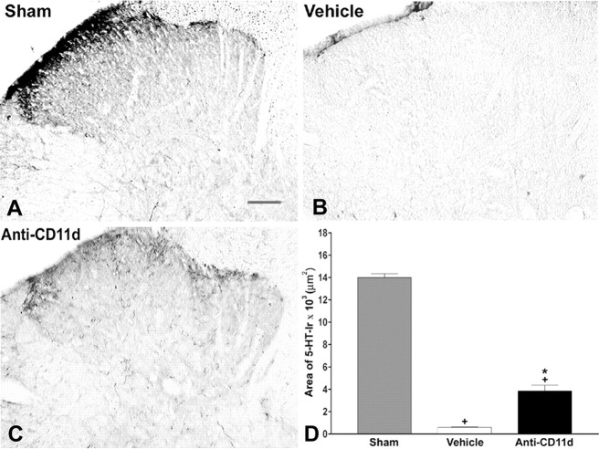
Figure 2.
5-HT-IR in laminas I-IV of the dorsal horn caudal to injury 4 weeks after SCI. Photomicrographs of transverse sections of the dorsal horn in sham-injured, vehicle-treated, and anti-CD11d mAb-treated animals. After SCI, 5-HT-immunoreactive fibers were completely absent in most dorsal horn sections. 5-HT-immunoreactive fibers were present as tortuous fiber within the superficial laminas after anti-CD11d mAb treatment. Scale bar, 100 μm. The area of 5-HT-IR (mean ± SE) in the dorsal horn caudal to the injury at T12-13 is plotted for the sham-injured (n = 5), vehicle-treated (n = 5), and anti-CD11d mAb-treated (n = 5) groups. *p < 0.05 compared with vehicle-treated rats; +p < 0.05 compared with sham-injured rats.

