
Figure 7.
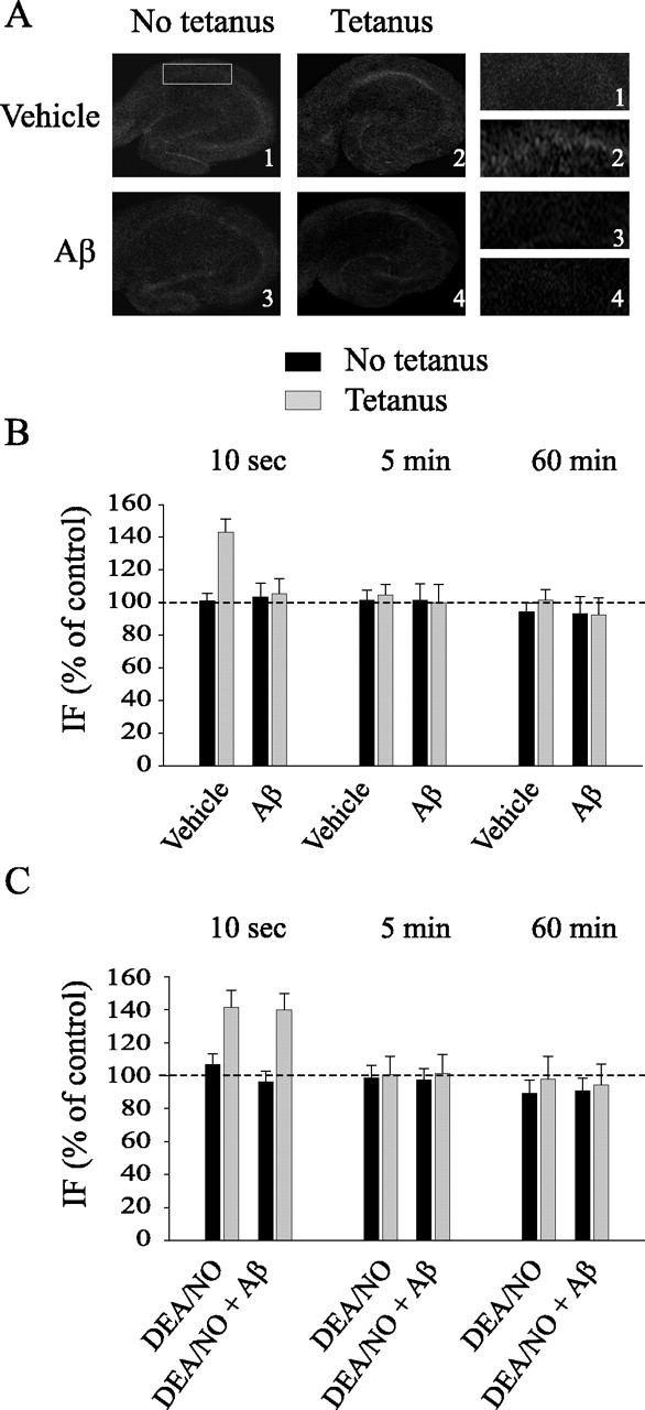
Aβ blocks the increase in cGMP immunofluorescence occurring immediately after LTP. A, Representative examples of hippocampal slices stained with a cGMP antibody and fixed 10 s after either vehicle plus tetanus and Aβ or Aβ plus tetanus. Left, Lower-power (4×) view of the entire slice. Right, Higher-power (16×) view of CA1 cell pyramidal area. B, Hippocampal slices are fixed 10 s, 5 min, or 60 min after vehicle or Aβ both in basal conditions and after tetanus. Aβ blocks the increase of cGMP immunofluorescence in tetanus-treated slices at 10 s (t(7) = 0.40; p > 0.5 between vehicle-treated slices and slices treated with Aβ plus tetanus). cGMP immunofluorescence does not vary at 5 or 60 min after the tetanus (t(6) = 0.25; t(6) = 0.19; p > 0.5). C, DEA/NO reestablishes the increase of cGMP immunofluorescence in tetanized slices exposed to Aβ at 10 s (t(8) = 0.022; p > 0.5 compared with tetanized slices). cGMP immunofluorescence does not vary at 5 or 60 min after the tetanus (t(6) = 0.30; t(6) = 0.33; p > 0.5). IF, Immunofluorescence. Error bars represent SEM.