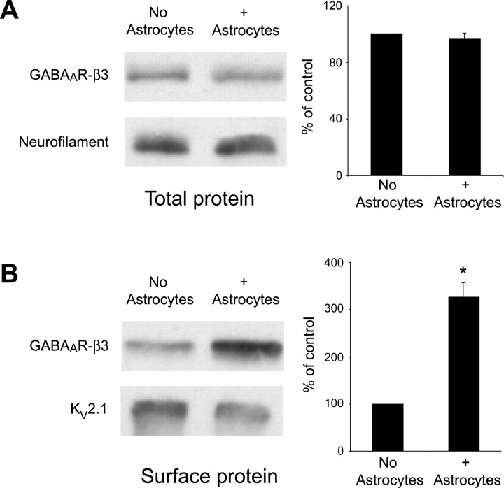
Figure 3.
Surface GABAAR expression is increased in hippocampal neurons in the presence of astrocytes. Total cell lysates or biotinylated surface protein extracts were harvested from hippocampal neurons cultured in the presence and absence of astrocytes at 10 div. A, Western blot analysis on total cell homogenates was performed using an antibody against the GABAAR-β3 subunit (top) or neurofilament H (bottom) as a loading control. Quantification of relative band intensity compared with loading controls demonstrates no significant difference in the level of GABAAR-β3 expression in the presence of astrocytes. B, Western blot analysis on surface-biotinylated extracts was performed using an antibody against the GABAAR-β3 subunit (top) or Kv2.1 (bottom) as a loading control. Quantification of relative band intensity compared with loading controls shows that GABAAR-β3 expression at the neuronal surface is increased ∼3.5-fold when neurons are cultured in the presence of astrocytes (*p < 0.001). Error bars represent SEM.

