Figure 4.
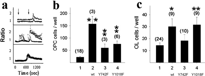
Rescue of PDGFRα-null OPCs with wild-type and signaling uncoupled receptor transgenes. a, Point mutations uncouple PDGFRα from specific second-messenger pathways. Intracellular free calcium in control OPCs (top), OPCs expressing wild-type receptor transgenes (middle), and OPCs expressing mutant receptor transgenes defective for PLCγ activation (bottom). The analysis used chimeric fms:pdgfra transgenes, and tracings represent the emission ratio (560/520 nm) from individual fura-2-loaded cells (>40 cells imaged per field) sequentially stimulated with the Fms-specific ligand hCSF and then with PDGF-AA to activate endogenous PDGFRα. Vertical arrows in the top panel denote the time of stimulation, and an absence of hCSF response in the bottom panel identifies PDGFRα tyrosine 1018 as essential for PLCγ activation. b, c, Impaired rescue of OPCs and oligodendrocytes (OL) with point-mutant receptor transgenes. Mean ± SEM number of O4+ OPCs (b) and MBP+ oligodendrocytes (c) per well in PDGFRα-null spinal cord explant cultures (1) and parallel cultures after transfection with expression vectors encoding wild-type (wt) PDGFRα (2), PDGFRαY742F (3), or PDGFRαY1018F (4). Values in parentheses represent the number of independent experiments, and asterisks indicate values that are statistically significant from nontransfected control cultures (*p < 0.05; **p < 0.01).

