
Figure 2.
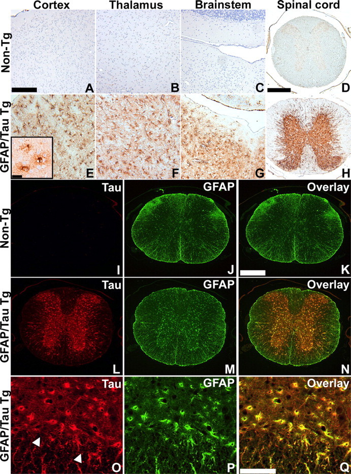
Astrocyte-specific regional tau expression in GFAP/tau Tg mice. A-H, Immunohistochemistry was performed with the human tau-specific MAb OT12 on 6-month-old non-Tg (A-D) and GFAP/tau Tg line 4 (E-H) mice from the CNS regions as indicated. Consistent with the biochemical analysis (Fig. 1), the highest levels of expression were observed consistently in the gray matter of the spinal cord. The inset in E shows morphology of cortical astrocytes at high magnification. Scale bars: (in A) A-C, E-G, 200 μm; (in D) D, H, 400 μm; E, inset, 25 μm. I-Q, Spinal cord sections from 6-month-old non-Tg (I-K) and GFAP/tau Tg, line 4 (L-Q) mice were double-labeled with OT12 (green; I, L, O) and GFAP (red; J, M, P). Merged images are depicted in K, N, and Q. Prominent GFAP staining is observed in astrocytes within the gray matter of Tg mice that is not detected in non-Tg animals. Arrowheads in O indicate the junction of the gray and white matter in the anterior horn of the spinal cord. Scale bars: (in K) I-N, 400 μm; (in Q) O-Q, 100 μm.