
Figure 3.
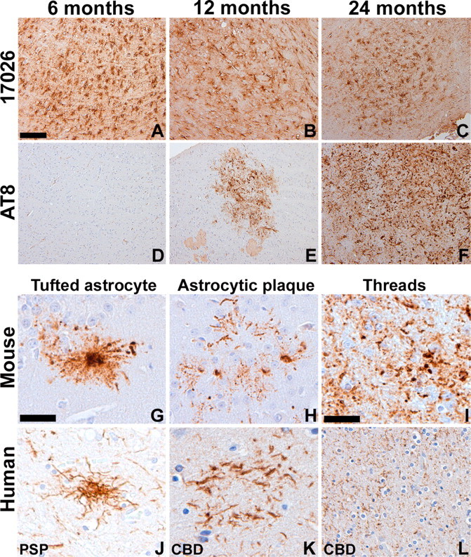
Accumulation of tau pathology in aged GFAP/tau Tg mice that resembles astrocytic pathology in human tauopathies. A-F, Immunohistochemical analysis was performed on sections of medulla from 6-month-old (A, D), 12-month-old (B, E), and 24-month-old (C, F) GFAP/tau Tg mice, line 4, as indicated with antibodies to recombinant tau (17026; A-C) or tau phosphorylated at Ser202 and Thr205 (AT8; D-F). There is an age-dependent accumulation of phosphorylated tau epitopes detected in GFAP/tau Tg mice. G-L, High-power photomicrographs of astrocytic pathology of Tg mice (G-I) and human tauopathies (J-L). The astrocytic pathology in the Tg mice resembles the tufted astrocytes (G, J), astrocytic plaques (H, K), and thread pathology (I, L) observed in tauopathies such as PSP and CBD. Scale bars: (in A) A-F, 200 μm; (in G) G, H, J, K, 40 μm; (in I) I, L, 80 μm.