
Figure 4.
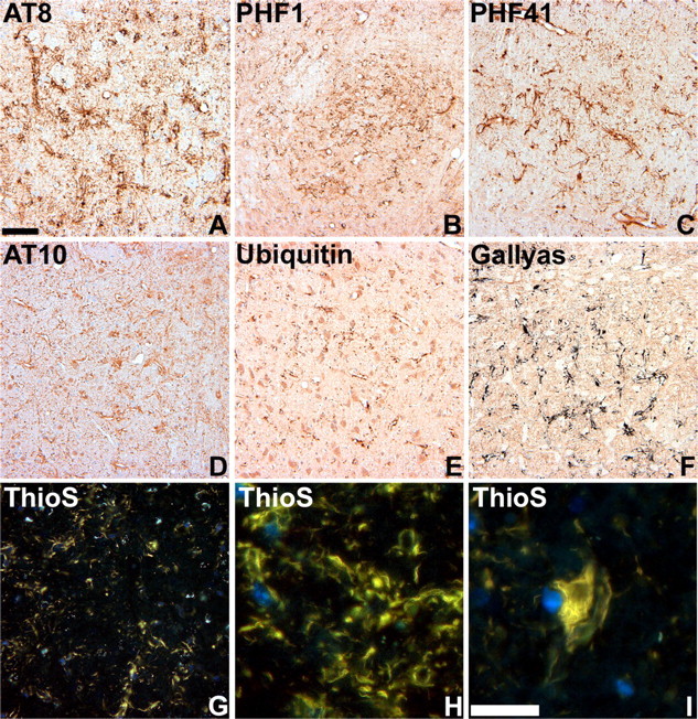
Characterization of tau pathology in GFAP/tau Tg mice. A-E, Immunohistochemical analysis was performed on sections of pons (A, B) and thalamus (C-E) from 22-month-old GFAP/tau Tg mice, line 4 with a panel of antibodies to distinct phosphorylated tau epitopes (A-D) and ubiquitin, as indicated. Ubiquitin (E) is detected only in a subset of tau pathologies. F-I, Gallyas silver impregnation (F) and thioflavine S (ThioS) (G-I) staining of astrocytic tau pathology in brainstem of 22-month-old GFAP/tau Tg mice. There is robust Gallyas-positive tau pathology in both astrocytic processes and cell soma. The thioflavine S stain detects only a subset of the pathology observed with antibodies to tau phosphoepitopes or Gallyas stains. Scale bars: (in A) A-G, 80 μm; (in I) H, I, 25 μm.