
Figure 2.
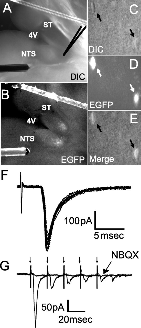
Solitary tract-evoked NTS synaptic responses in NTS POMC-EGFP neurons. A, B, Visualization of the NTS brain-slice preparation using both DIC (A) and fluorescence (B). Orientation of the mouse brainstem slices in the horizontal plane allowed the placement of the concentric bipolar stimulating electrode on the ST several millimeters from the recording region (dark gray center) in the medial NTS. 4V, Fourth ventricle. C, E, Identification of individual POMC-EGFP neurons by DIC (C), fluorescence (D), and merge (E). Two POMC-EGFP-positive neurons can be seen (arrows). F, ST activation evoked monosynaptic EPSCs in an NTS POMC-EGFP neuron; a total of 10 successive EPSCs are shown. Vm = -60 mV. ST shock evoked a short-latency EPSC with high reliability (latency, 1.58 ms; jitter, 65 μs and no observed failures at 50 Hz ST stimulation). G, Successive shocks (train of 5 pulses at 50 Hz; arrows) evoked a frequency-dependent depression of EPSC amplitude. The non-NMDA glutamate receptor antagonist NBQX (10 μm) completely blocked the EPSCs.