
Figure 3.
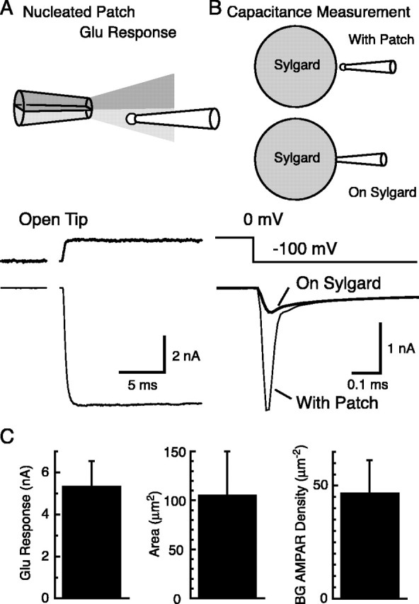
Density of AMPA receptors at the cell soma of BGs. A, Nucleated outside-out patches were taken from the soma of BGs by applying slight negative pressure inside the pipette while pulling the pipette away from the cell soma. Rapid application of 10 mm glutamate in the presence of 200 μm CTZ (top trace, open tip junctional current) resulted in large AMPA receptor-mediated currents (bottom trace). Vh = -70 mV. B, The same nucleated outside-out patches used for recording glutamate responses were used for capacitance measurements. Voltage steps of -100 mV were applied, and the resulting current responses were recorded (thin line, with patch). After touching the surface of a Sylgard bead, the capacitive current attributable to the patch disappeared (thick line, on Sylgard). The difference between the two currents was taken. Step response of the recording system to the same voltage pulses were determined by recording the response of a resistor connected to the head-stage of the patch amplifier. This trace was fitted to the final level of the leakage component. Subtraction of this leakage component leaves the capacitive current across the membrane patch. The membrane charge induced by the voltage pulse is given by the integration of this capacitive current (90.7 fC for this patch). C, Left, Summary of glutamate response amplitudes from nucleated outside-out patches. Middle, Area of the membrane calculated from the capacitive charge. Right, BG AMPA receptor (AMPAR) density calculated by dividing glutamate response amplitude by the membrane potential, POmax, and single-channel current of BG AMPA receptors (n = 6 patches). Error bars represent SD.