
Figure 2.
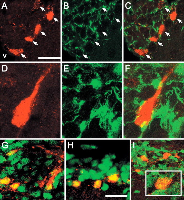
RA-activated SVZ cells express neural precursor and mitotic markers. A, B, Color-separated images of five adjacent transgene-expressing cells (A, arrows) situated within a network of nestin-labeled processes (B, arrows) in a single optical section (0.5 μm) through the SVZ, in a region adjacent to the ventricle (V). C, Merged image of A and B confirms coincidence of transgene and nestin labeling. D, E, Color-separated images of a fusiform transgene-labeled cell (D) with a single process oriented away from the ventricle seen within a network of nestin-labeled processes (E). F, Merged image of the single optical section in D and E shows that this cell, which has the morphology suggested for neural stem cells in the SVZ (Garcia et al., 2004) is double labeled for the transgene and nestin. G, A subset of Sox1-labeled SVZ cells (green) also expresses the transgene (red). H, A subset of Sox2-labeled cells (green) also expresses the transgene(red). I, Transgene-labeled cells (red) express p27Kip1 (green), which is more broadly expressed in the adult SVZ. Inset, An example of a single large transgene-labeled cell that also expresses p27Kip1. Scale bars: A-C, 10 μm; G, H, 10 μm; width of images in D-F, 10 μm.