
Figure 5.
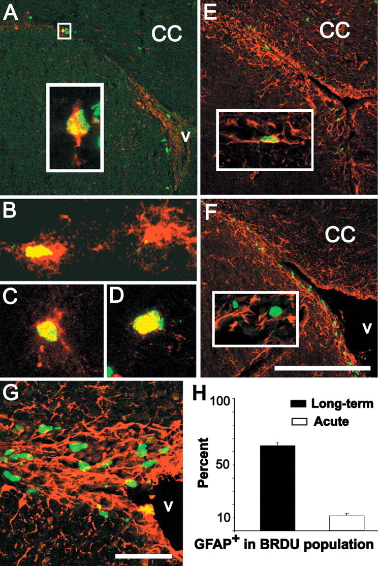
RA-activated cells in the SVZ are a subset of resident, slowly dividing precursors. A, Transgene (red) and BrdU-labeled cells (green) in the SVZ (coronal section) 30 d after long-term exposure (10 mg/ml for 1 week in the drinking water). Inset, High magnification of an optical section (0.5 μm) showing a large transgene-labeled SVZ cell (A, box) with as single process oriented away from the ventricle that is also labeled by BrdU 30 d after a long-term exposure. B-D, Examples of transgene-labeled SVZ cells that are also labeled after long-term BrdU exposure (yellow nuclei in cells with red processes and cytoplasm). E, Thirty days after long-term exposure, several BrdU-labeled cells are scattered throughout the CC but, as shown in A, are rare in the SVZ. Inset, A long-term BrdU-labeled SVZ cell is clearly labeled for GFAP. F, Distribution of BrdU- and GFAP-labeled SVZ cells after acute BrdU exposure (2.5 h). The BrdU-labeled cells are more frequent. Inset, Most acutely labeled BrdU-positive cells do not express GFAP. G, Example of the 40× confocal maximum projection images used to estimate numbers of BrdU-labeled cells that express GFAP after acute exposure. H, Quantitative estimate of the proportion of long-term versus acute BrdU-labeled cells that also express GFAP. Scale bars: A, E, F, 200 μm; G, 25 μm; width of image in B, 50 μm; width of images in C, D, 25 μm.