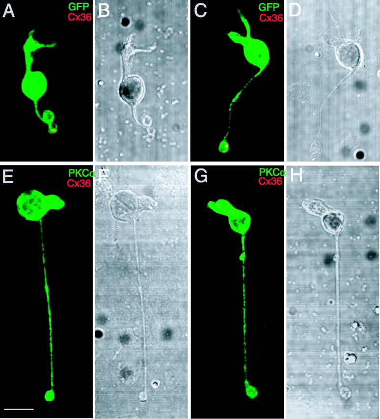
Figure 2.

Expression of Cx36 in isolated 357 bipolars, not in isolated rod bipolars. A-D, Two isolated 357 bipolars labeled with the Cx36 antibody. A, C, Axon terminals of the cells (green) colocalized with Cx36-immunoreactive punctate (red). B, D, DIC images of the two same cone bipolar cells. E-H, Two isolated rod bipolars double labeled with PKCα and Cx36 antibodies. E, G, No Cx36-immunoreactive puncta colocalize with the rod bipolars (green). F, H, DIC images of the two same rod bipolars. Scale bar, 5 μm.
