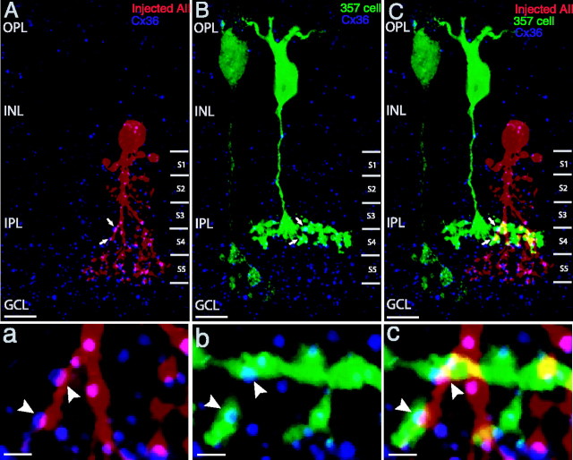
Figure 4.
Cx36 appears to be present at contacts of a 357 bipolar with AII amacrine cells. A-C, Confocal projected images. Cx36 (blue) is seen in the dendrites of an injected AII amacrine cell (red, A) and in the axonal terminals of a 357 bipolar (green, B). A, B, Some of the Cx36-immunoreactive puncta colocalize with both (arrows). C, The merged image of A and B demonstrates that the Cx36 puncta appear at contacts of the 357 bipolar with AII amacrine cell (arrows). a-c, High-magnification views of single optical sections show Cx36 puncta occur at the gap junctions of the 357 bipolar with AII amacrine cell (arrowheads; c) and colocalize with dendrites (arrowheads; a) and axonal terminals (arrowheads; b). Scale bars: A-C, 10 μm; a-c, 2 μm. OPL, Outer plexiform layer; INL, inner nuclear layer; IPL, inner plexiform layer; GCL, ganglion cell layer.

