
Figure 5.
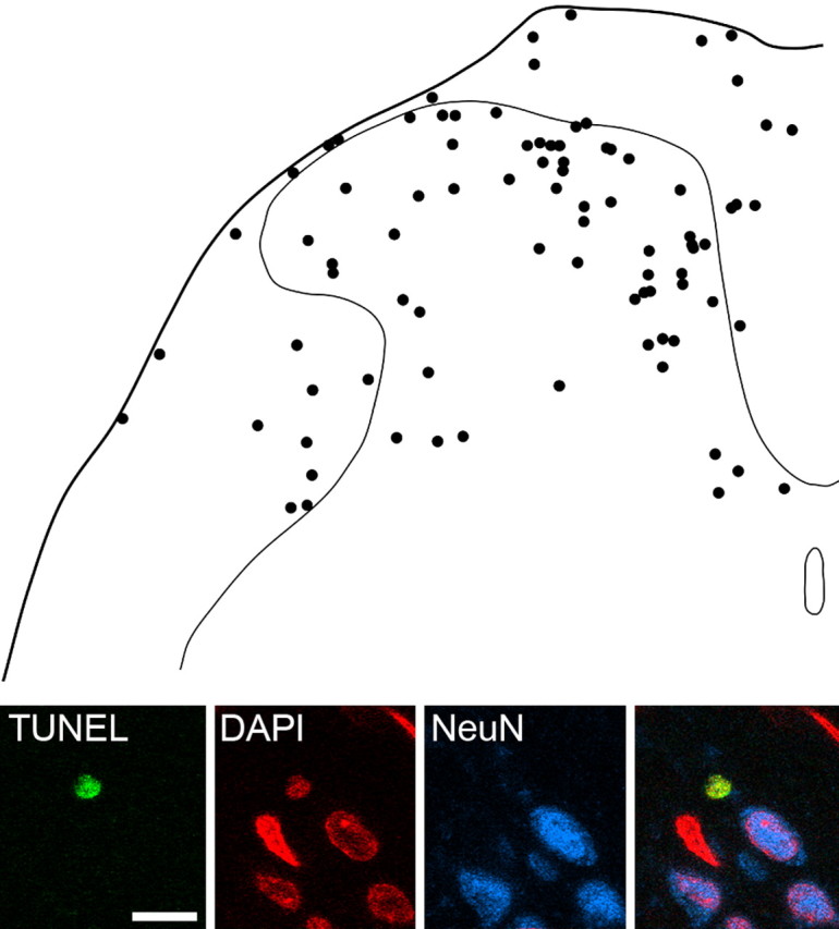
TUNEL staining in the dorsal spinal cord 7 d after SNI and lack of coexistence with NeuN immunoreactivity. The drawing shows the positions of 86 TUNEL-positive nuclei that were present on the side ipsilateral to the SNI in five sections each from six rats, plotted onto an outline of the dorsal half of the spinal cord. Note that although the majority of TUNEL-positive nuclei are in the gray matter, there are also many throughout the white matter. The confocal images in the bottom part of the figure show a single optical section through a TUNEL-labeled nucleus in lamina I that is not NeuN immunoreactive. Other NeuN-positive and NeuN-negative nuclei are also visible. Scale bar for confocal images, 10 μm.