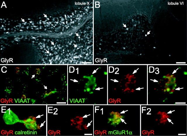
Figure 5.
Expression of GlyRs by UBCs. A, B, Immunodetection of GlyR in lobules X and VI, respectively. Numerous GlyR-IR structures (arrows) within lobule X granular layer contrasting with small numbers of GlyR-IR profiles (arrows) in lobule VI. C, F, Double detection of GlyR and different markers in the granular layer of lobule X. C, Codetection of VIAAT (green) and GlyR (red) indicating that only a subpopulation of glomeruli expresses GlyR-IR (arrows). D, High magnification of a glomerulus with presynaptic VIAAT-IR (green) apposed (arrows) to postsynaptic GlyR clusters (red). E, GlyR aggregates (red, arrows) on the dendritic brush of a CR-positive UBC (green). F, GlyR aggregates (red, arrows) are also detected on mGluR1α-IR (green) UBC dendrioles. A-F, Wide-field CCD camera images. Scale bars: A, B, 100 μm; C, 20 μm; D-F, 5 μm.

