
Figure 1.
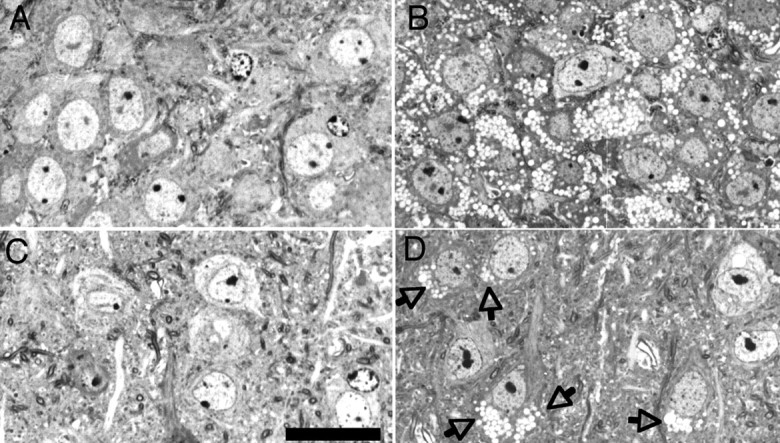
Light micrographs demonstrate lysosomal storage in the CNS of α-mannosidase-deficient mice. Micrographs show the hippocampal CA3 region (A, B) and lamina 5/6 neocortex (C, D) in 2-month-old wild-type (A, C) and α-mannosidase-deficient (B, D) mice. In the hippocampal CA3 region (B), most neuronal perikarya show clear cytoplasmic vacuoles that are the morphological equivalent for lysosomal storage of mannose-rich oligosaccharides. In the neocortex (D), vacuolation of neuronal perikarya (arrows) is more variable. Semithin sections, Toluidine blue staining. Scale bar, 20 μm.