Figure 1.

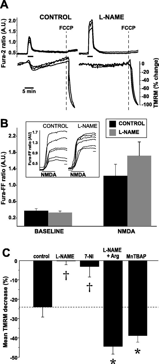
Inhibition of NOS prevents NMDA-induced ΔΨm dissipation. A, Simultaneous measures over time of NMDA-induced changes in [Ca2+]i (fura-2) and ΔΨm (TMRM) in the absence (left) and presence (right) of l-NAME (100 μm for 3 h). Maximal ΔΨm dissipation is induced with FCCP (dotted line) at the end of each experiment. To eliminate contributions to changes in mitochondrial TMRM intensity made by plasma membrane depolarization, TMRM intensities from a mitochondria-rich region of each neuron at each point in time was divided by the corresponding intensity from a mitochondria-poor region within the same neuron. Each ratio was normalized to the mean baseline ratio (100%) and the mean ratio obtained after FCCP (0%). NMDA induces a modest ΔΨm dissipation that is blocked by l-NAME. B, Population summary of mean peak [Ca2+]i increases during NMDA in the presence and absence of NOS blockade. [Ca2+]i increases are reported with fura-FF (Kd of 5.5 μm). NMDA-induced [Ca2+]i increases do not significantly differ between control and l-NAME. Inset, Representative examples of raw fura-FF responses to NMDA in control and l-NAME-treated neurons. Calibration bar, 1 min. C, NMDA-induced ΔΨm dissipation is decreased during NOS blockade (l-NAME, 7-NI), and the blockade is overcome with supplemental l-arginine (Arg). MnTBAP, a superoxide dismutase mimetic, increases NMDA-induced ΔΨm dissipation, suggesting that a portion of the NO generated by NMDA is converted to peroxynitrite. Statistical comparisons made between control and each treatment. *p < 0.05; †p < 0.01. A.U., Arbitrary units.
