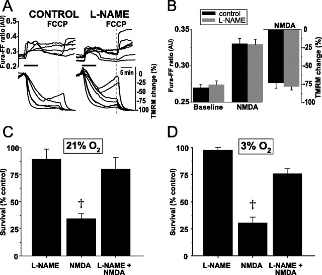
Figure 9.
In hippocampal neurons from P19 rats, NO production does not mediate the marked, NMDA-induced ΔΨm dissipation but does induce widespread neuronal death after NMDA. A, Simultaneous measures over time of NMDA-induced changes in [Ca2+]i (fura-FF, Kd of 5.5 μm) and ΔΨm (TMRM) in the absence (left) and presence (right) of l-NAME (100 μm for 3 h). Maximal ΔΨm dissipation is induced with FCCP (dotted line) at the end of each experiment. In contrast with P5 hippocampal neurons, NMDA induces profound ΔΨm dissipation. l-NAME fails to block this dissipation. B, Population summary of mean peak [Ca2+]i increases and ΔΨm dissipation during NMDA in the presence and absence of l-NAME. The magnitudes of neither the [Ca2+]i increases nor the ΔΨm dissipation differ significantly between control and l-NAME-treated neurons. C, NO mediates neuronal death in P19 neurons from NMDA in 21% O2. Survival was quantified by live-dead assay 48 h after a 20 min exposure to NMDA (300 μm for 20 min) and is expressed as a percentage of survival after exposure to control, bicarbonate-buffered saline alone (see Materials and Methods). l-NAME incubation alone does not alter survival. NMDA decreases survival to 25% of control. l-NAME pretreatment prevents the majority of NMDA-induced death. D, When applied in 3% O2, NMDA induces the same degree of neuronal death seen in 21% O2. The marked neuroprotection conferred by l-NAME pretreatment before NMDA exposure in 3% O2 demonstrates that physiologic O2 tension does not affect the neurotoxic effects of NMDA-induced NO production. †p < 0.01.

