Figure 5.

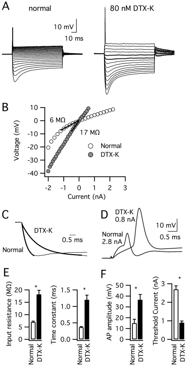
The application of DTX-K altered the electrophysiological properties of MSO principal neurons exhibiting mature features (P20–P21). A, The response of a typical MSO neuron to current pulses (–2 to 0.8 or 2.8 nA, 0.2 nA steps) before and after the application of 80 nm DTX-K shows that a transient firing pattern persisted in the presence of the toxin. B, In contrast, the slope of the peak V–I plot below rest reveals that the input resistance of this cell increased >2.5-fold. C, Normalized inward voltage responses show that the neuron's membrane time constant also increased threefold from 0.4 to 1.3 ms after toxin application. D, Action potentials in this neuron increased in amplitude and the threshold current for action potential initiation decreased in response to DTX-K. E, F, Average membrane time constant, peak input resistance, threshold current for action potential initiation and action potential amplitude before (open bars) and after (filled bars) DTX-K application (n = 5). All parameters were significantly different in DTX-K (*p < 0.001). AP, Action potential. Error bars represent SEM.
