
Figure 6.
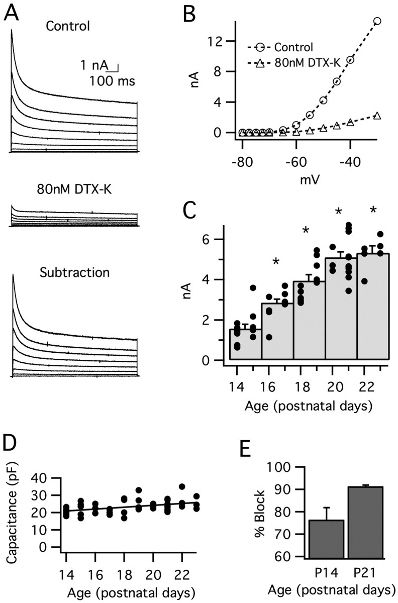
Low voltage-activated potassium current (IK(LVA)) increases after hearing onset in MSO principal neurons. IK(LVA) was isolated pharmacologically (see Materials and Methods) and evoked by a family of 1 s depolarizations (–70 to –30 mV; 5 mV steps; from a prepulse potential of –80 mV). A, In a typical neuron from P21, IK(LVA) (top) was essentially blocked by the addition of 80 nm DTX-K (middle). DTX-K-sensitive currents are shown in the bottom panel. Traces are not leak-subtracted. B, Peak I–V curves for leak-subtracted IK(LVA) before (circles) and after (triangles) the addition of 80 nm DTX-K (the same neuron as A). For this neuron, IK(LVA) was blocked by 80–90% across the range of voltages shown. C, Peak IK(LVA) (leak-subtracted) as a function of age. This current increases 390% between P14 and P23. The histogram displays peak current responses for voltage steps to –45 mV, grouped in 2 d bins. The superimposed filled circles represent currents from individual cells. The asterisks indicate significant difference from the P14–P15 group; p < 0.001. D, Whole-cell capacitance did not change significantly between P14 and P23, indicating that changes in cell size/membrane area do not account for the developmental increase in IK(LVA). E, DTX-K (80 nm) blocks the majority of IK(LVA) in response to steps to –45 mV. The percentage block increases from 76 to 91% between P14 and P21 (p = 0.06; n = 4–6). Error bars represent SEM.