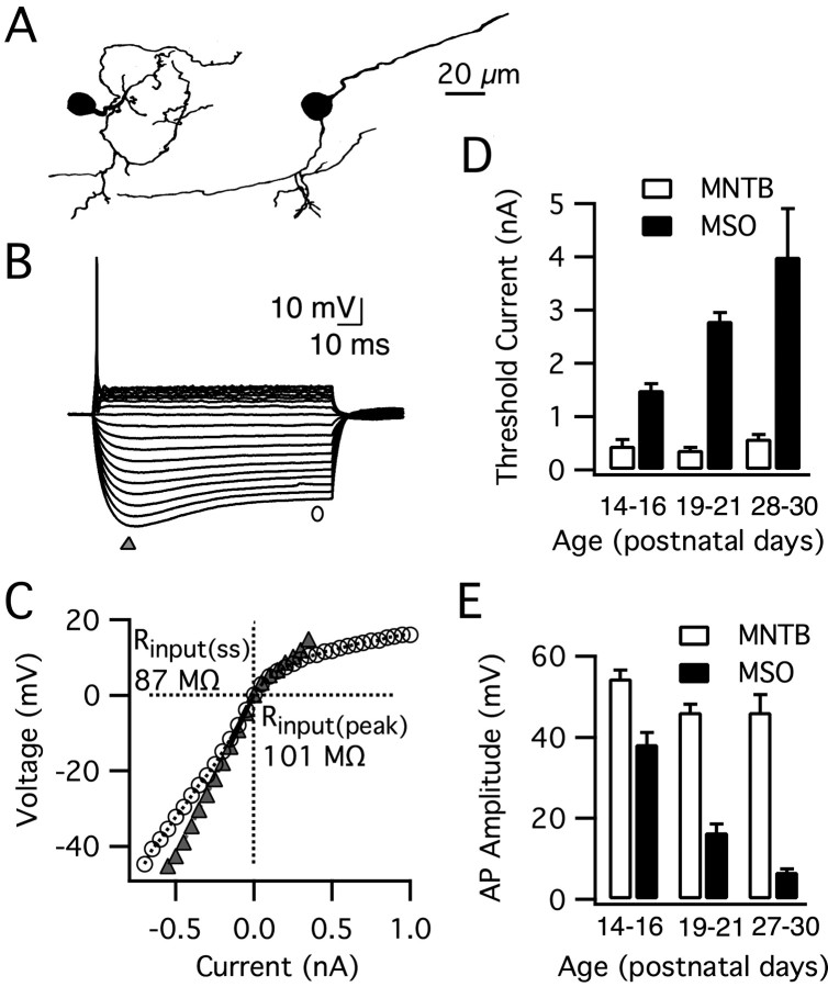
Figure 8.
Principal neurons of the MNTB do not undergo changes in their intrinsic properties after hearing onset. A, Camera lucida tracings of two biocytin-labeled MNTB principal neurons show their characteristic ovoid somata and small-caliber dendrites. B, Responses to step pulses (–0.55 to 0.35 nA, 0.05 nA steps) for a typical MNTB principal cell (P15; resting membrane potential, –68 mV). MNTB principal neurons exhibit a transient firing pattern with strong outward rectification, similar to MSO principal neurons (see Fig. 1 A). C, The peak (triangle) and steady-state (circle) V–I plot for this MNTB principal neuron demonstrates that the input resistance near rest of MNTB neurons is larger than MSO neurons. The input resistance was obtained from the slope of the V–I plot between 0 and 10 mV below rest (Rinput(pk) = 101 MΩ, Rinput(ss) = 87 MΩ). D, E, MNTB principal cells fired large action potentials 2–4 weeks after birth with no significant change in the threshold current for initiation (p > 0.05; n = 6–7) or amplitude (p > 0.1; n = 6–7). In contrast, the action potential signaling properties of MSO principal neurons changed dramatically (threshold current, p < 0.001; amplitude, p < 0.001; n = 7–21, n = 7–20). Overall, MNTB principal neurons required far more modest currents for action potential initiation and had larger action potentials relative to principal neurons of the MSO (threshold, p < 0.001, n = 6–20; amplitude, p < 0.001, n = 6–21).

