
Figure 3.
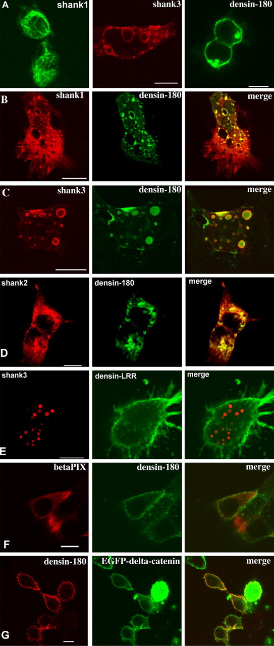
Shank targets Densin-180 into intracellular clusters. Expression vectors for EGFP-shank1 (A), Shank1-3 without tag (B-G), βPIX, EGFP-δ-catenin, and Densin-180 were transfected into HEK cells either alone (A) or in combination (B-G). Shank was visualized by staining using EGFP autofluorescence (A) or with rbAnti-shank, followed by Cy3-labeled anti-rabbit antibodies (red fluorescence). Densin-180 was visualized by monoclonal mouse anti-myc antibody, followed by Cy2-labeled anti-mouse antibodies (A-C, green fluorescence), by rbAnti-Densin-180, followed by Cy2-labeled anti-rabbit (D, red fluorescence), or by mouse anti-myc, followed by Cy3-labeled anti-mouse (E, red fluorescence). βPIX was labeled by anti-T7, followed by Cy3-labeled anti-mouse (D, red fluorescence), and δ-catenin was visualized by the EGFP-autofluorescence (E). All pictures are confocal sections taken approximately at the level of the center of the cells. Scale bars, 5 μm.