Figure 7.
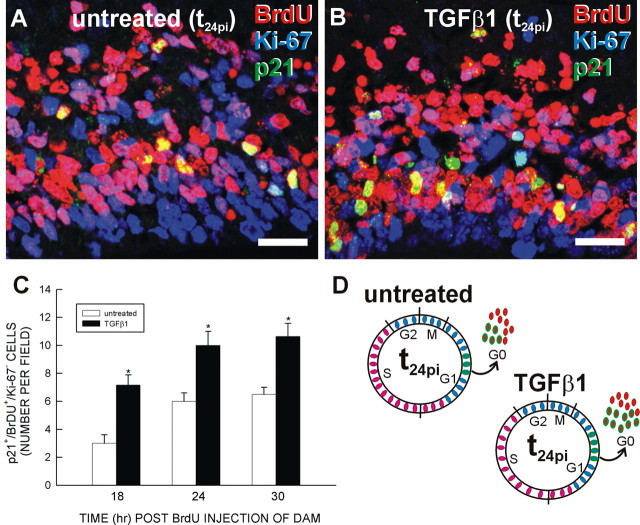
TGFβ1 increases p21-mediated cell cycle exit. After collection at 1 h after BrdU administration (t1pi), slices were treated with TGFβ1 (0 or 40 ng/ml), collected at t18pi, t24pi, and t30pi, and triple immunolabeled for p21, BrdU, and Ki-67. A, B, Qualitatively, the number of p21+, BrdU+, and Ki-67– cells at t24pi was increased in the slice treated with 40 ng/ml TGFβ1 compared with untreated. C, The number of p21+, BrdU+, and Ki-67– cells in a field was determined for untreated and TGFβ1-treated slices. At each time point, TGFβ1 increased the number of p21+, BrdU+, and Ki-67– cells. D, The diagram illustrates the effect of TGFβ1 on p21-mediated cell cycle exit. Asterisks indicate a statistically significant (p < 0.05) difference from untreated. Scale bars, 20 μm.

