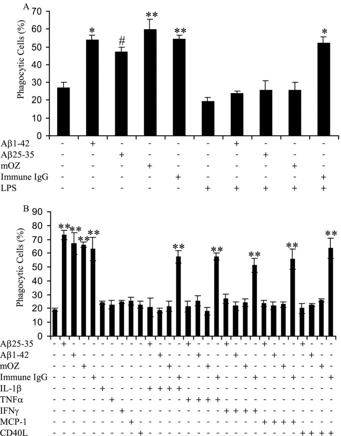
Figure 1.
Proinflammatory cytokines inhibit fAβ and opsonized zymosan-stimulated phagocytosis but do not affect immune IgG phagocytosis. A, BV-2 cells were exposed to LPS (1μg/ml) for 6 h to produce a proinflammatory environment. The cells were then stimulated with fAβ (60 μm fAβ25–35 and 5 μm fAβ1–42), mouse complement-opsonized zymosan (mOZ; 1 mg/ml), and immune IgG (1 mg/ml) for 30 min, followed by 30 min of incubation with fluorescent microspheres. The fraction of phagocytically active cells was then determined. B, BV-2 cells were incubated with proinflammatory cytokines (15 ng/ml IL-1β, 10 ng/ml TNFα, and 100 ng/ml IFNγ), CD40L (1 μg/ml), or MCP-1 (20 ng/ml) overnight before stimulation with phagocytic-stimulating ligands for 30 min. Fluorescent microspheres were then added to the cells for 30 min. The fraction of phagocytically active cells was then determined. *p < 0.01, **p < 0.001, and #p < 0.05 compared with control. This experiment is representative of three independent experiments.

