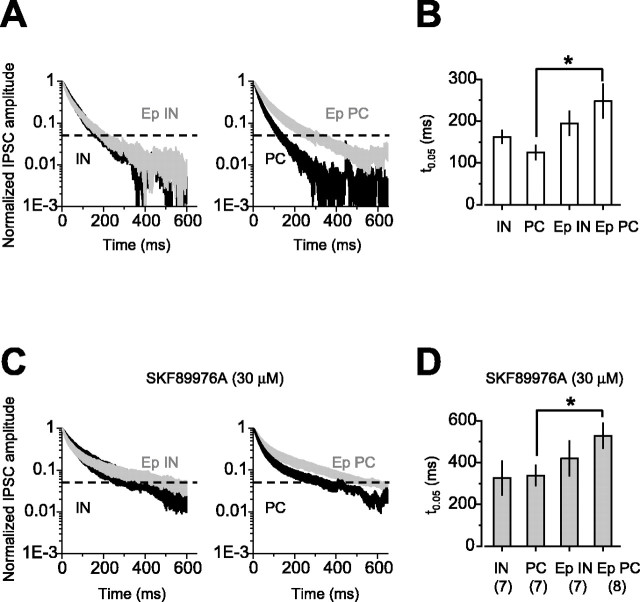
Figure 3.
The late component of eIPSCs increases in pyramidal cells after status epilepticus. A, Average of the decaying phase of eIPSCs normalized by their peak amplitude in interneurons and pyramidal cells from control (black traces; IN, n = 7; PC, n = 7) and epileptic (gray traces; Ep IN, n = 7; Ep PC, n = 8) rats. eIPSCs in pyramidal cells from epileptic animals had a more prolonged time course than in pyramidal cells from control rats. B, Summary data of the time at which the amplitude of the eIPSCs had decayed to 5% of the peak amplitude (t(0.05)). After epilepsy, eIPSCs recorded from pyramidal cells had a slower time course. C, Averaged decaying phases of eIPSCs recorded from the same cells as in A in the presence of SKF89976A (30 μm). D, Summary data of t(0.05) recorded in the presence of SKF89976A. Blocking GABA transporters prolonged the decay of eIPSCs to a similar extent in all the cell types. Even in the presence of SKF89976A, the decay was longer in pyramidal cells from epileptic animals than in pyramidal cells from control rats. *p < 0.05. Error bars represent SEM.

