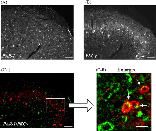
Figure 4.
A, B, Colocalization of PAR-1 (A) with PKCγ, which is highly limited to the inner part of lamina II in the dorsal horn of the mouse spinal cord (B). C-i, C-ii, The green labeling for PAR-1 and the red labeling for PKCγ show colocalization in the dorsal horn of the spinal cord. PAR-1-like IR is seen in the membrane of PKCγ-labeled cells in the dorsal horn of the spinal cord (arrowhead in C-ii). Scale bars: A, B, C-i, 50 μm; C-ii, 10 μm.

