
Figure 2.
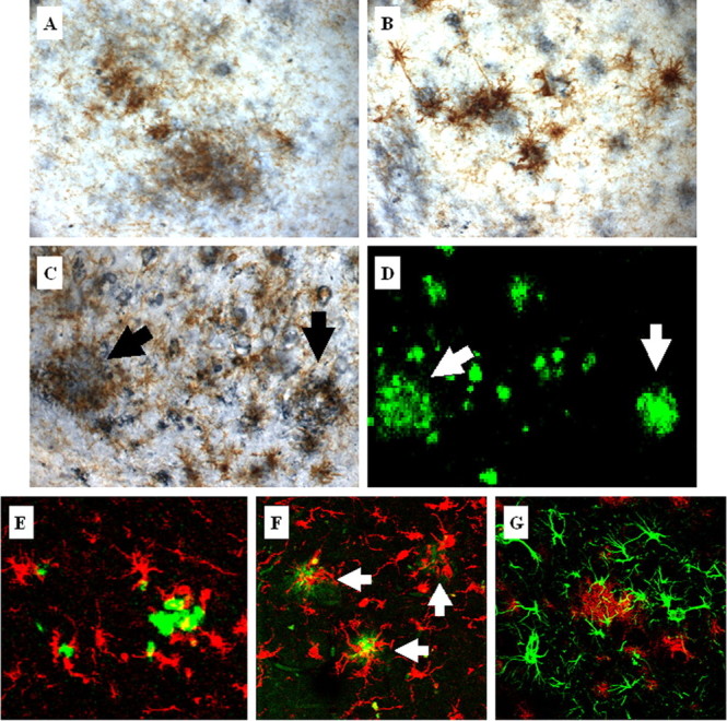
Activated microglia colocalize with plaques in aged 3xTg-AD mice. Activated microglia detected by CD45 immunoreactivity (brown) colocalize with extraneuronal Aβ-immunopositive deposits (blue) in the cortex (A), amygdala (B), and hippocampus (C) in 24-month-old 3xTg-AD mice. Serial brain sections of the hippocampus were stained with thioflavin S (D). Arrows point to activated microglia around thioflavin S-positive plaques. Additional double-staining fluorescent analysis using thioflavin S (green) and Iba1 (red) to detect microglia demonstrates that 3xTg-AD mice (E) show similar microglia and plaque interactions as observed in the hippocampus of the human AD brain (F). Arrows point to microglia associated with the cores of amyloid plaques in the human AD brain. G, Double fluorescent labeling of GFAP-immunopositive astrocytes (green) and amyloid-containing plaques (red) detected by 6E10 in 24-month-old 3xTg-AD mice shows colocalization of astrocytes around plaques.