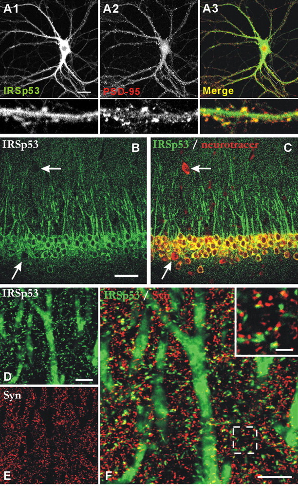
Figure 3.

Colocalization of IRSp53 with PSD-95 in cultured neurons and synaptic localization of IRSp53 in brain sections. A, Colocalization of IRSp53 and PSD-95 in cultured neurons. Cultured hippocampal neurons (21 DIV) were doubly stained by immunofluorescence for IRSp53 (A1; green) and PSD-95 (A2; red). The bottom panels are enlarged images of the small white boxes in A1, A2, and A3. B, C, Expression pattern of IRSp53 (B; green) in the CA1 region of rat hippocampus. The Neuro Tracer dye (C; red) was used to visualize all of the neurons. The arrows point to probable GABAergic interneurons. D-F, Colocalization of IRSp53 (D; green) with synaptophysin (E; Syn; red). The inset in the merged image (F) shows the close apposition or partial colocalization between IRSp53 and synaptophysin. Scale bars: A, 20 μm; B, 100 μm; D, F,10 μm; inset, 2 μm.
