
Figure 4.
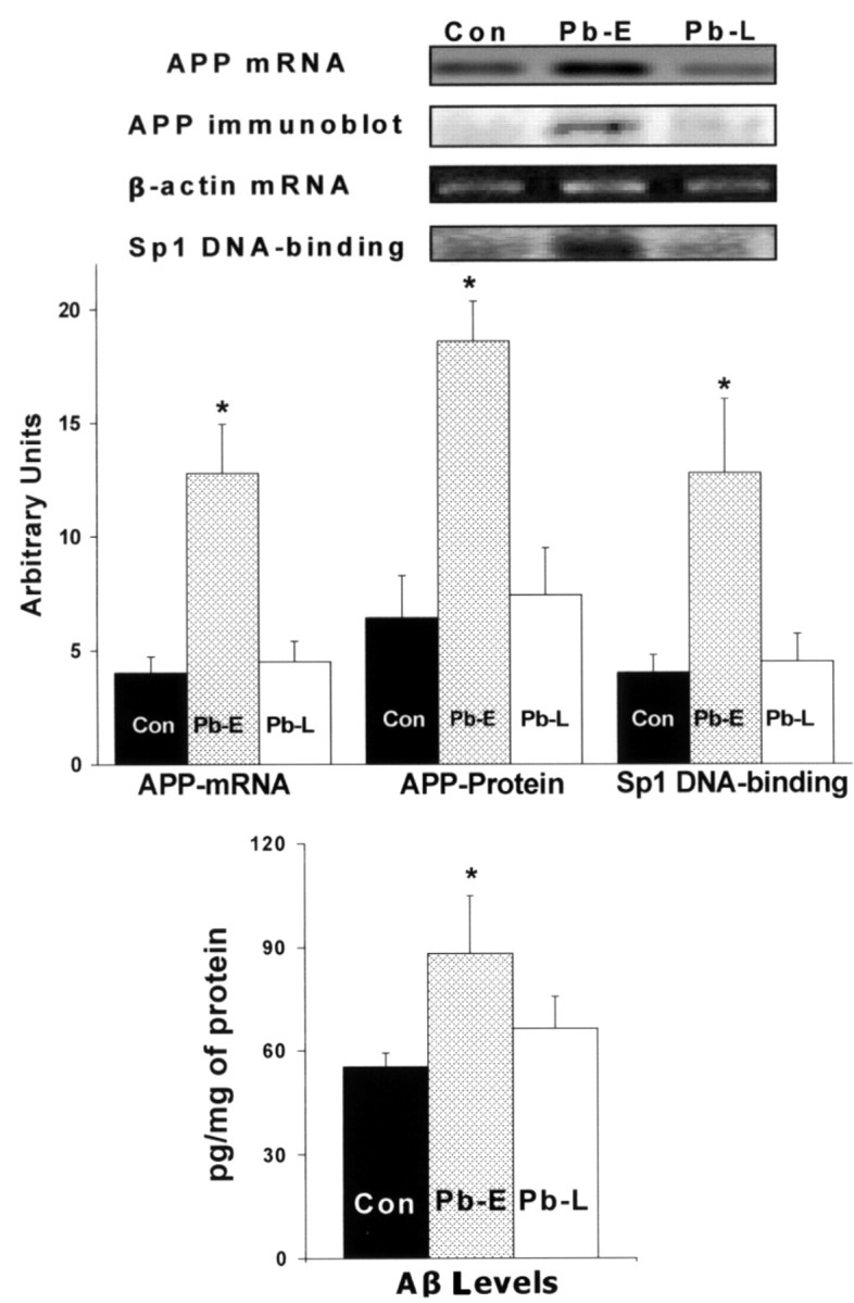
Unresponsiveness of APP expression, Sp1 DNA-binding, APP and Aβ formation to Pb exposure in old age. Brain cortices were obtained from control 20-month-old animals and those directly exposed to 200 ppm of Pb-acetate as follows: control (Con, unexposed), Pb-E (exposed from P1 to P20), and Pb-L (exposed from 18 to 20 months of age). These tissues were used to analyze the mRNA expression of APP, β-actin (RT-PCR), Sp1 DNA-binding (gel shift), APP protein levels (Western blot), and Aβ activity (ELISA). Data shown are derived from two cohorts of animals separated in time by 1 year; each data point represents the mean ± SEM for four to six animals. Data were analyzed by two-way ANOVA followed by a Duncan's post hoc test to compare the effects among various treatments; values marked with an asterisk are significantly different from their corresponding controls (p < 0.05).