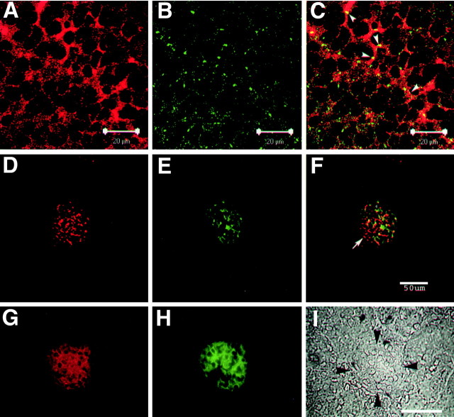
Figure 2.
NPCD colocalizes with PTPRO in brain and kidney. P0 mouse hippocampal sections (A-C) and adult kidney sections (D-F) were doubly immunostained with anti-Ptx (A,D) and anti-PTPRO (B,E), and confocal analysis (0.5 μm optical sections) was performed. Overlays are shown in C and F. Ptx-containing isoforms of NPCD appear localized mainly to the plasma membrane in CA3 hippocampal pyramidal neurons, where there is clear overlap (arrowheads, yellow; C) but not complete colocalization with PTPRO. Coexpression is present in the kidney, but there is extensive NPCD staining not associated with PTPRO (arrow, F). Conventional fluorescence microscopy of adult kidney sections (G-I) shows that both NPCD staining (red; G) and PTPRO staining (green; H) are confined to glomeruli (bordered by arrowheads in phase-contrast image; I) in adult kidney (Beltran etal., 2003). Scalebars: A-C,20 μm; F, I,50 μm.

