Figure 5.
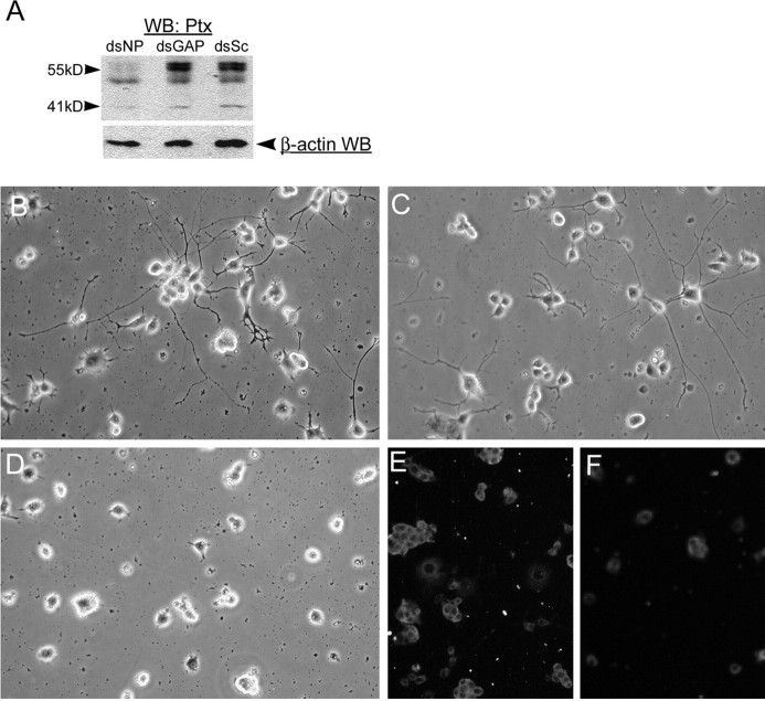
NPCD proteins are required for NGF-induced neuronal differentiation in PC12 cells. A, PC12 cells were transfected with siRNAs directed against NPCD (dsNP), GAPDH (dsGAP), or scrambled siRNAs (dsSc), and after 24 h, cell lysates were run on SDS-PAGE and sequentially blotted with anti-Ptx and anti-β-actin as a loading control. NPCD siRNAs severely reduced expression of the 55 kDa NPCD protein doublets (>90%) and caused a 40% reduction in expression of the 41 kDa NPCD protein compared with controls at this early time. B-F, PC12 cells were transfected with siRNAs that were scrambled so as not to match known sequences (B), siRNA capable of knocking down GAPDH (C, E), or siRNA for NPCD (D, F) and were then replated and grown in NGF for 3 d. Cells were examined by phase contrast (B-D) or fixed and stained for NPCD (E, F). Knock-down of NPCD was demonstrable after 4 d(F) and led to a lack of neurite growth (D). Hoechst staining of transfected cells revealed no apoptosis induced by NPCD siRNA (data not shown). These experiments were performed five times with similar results. Note that NPCD staining in PC12 cells requires permeabilization and is therefore cytosolic; the “ring” staining pattern suggests membrane association (Chen and Bixby, 2005). WB, Western blot.

