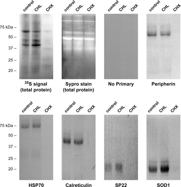
Figure 3.
Axoplasmic protein synthesis. Sheared axons were pretreated with 25 μg/ml chloramphenicol (CHL), 10 μg/ml cycloheximide (CHX), or vehicle (Control) for 20 min, and then 2 mCi/ml [35S]methionine/cysteine was added for 4 h. The first two panels show fractionated lysates from the axons with total protein visualized by Sypro Ruby stain (Sypro) and labeled proteins visualized by autoradiography (3 d exposure). The remainder of the panels show autoradiograms of immunoprecipitated proteins indicated (peripherin, ≈54 kDa; HSP70, ≈70 kDa; calreticulin, ≈48 kDa; Uch-L1, ≈25 kDa; SP22, ≈20 kDa; SOD1, ≈15 kDa). Note that newly synthesized proteins are detected in the control and CHL lanes but not in the CHX lane, whereas the Sypro stain of the lysates before immunoprecipitation shows approximately equivalent levels of unlabeled proteins in all lanes.

