Figure 4.
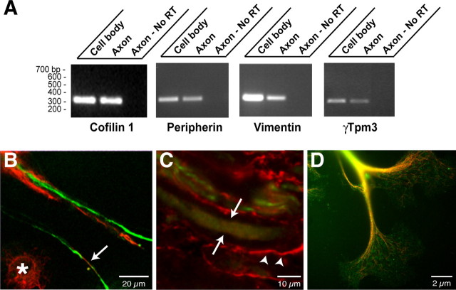
Cytoskeletal proteins and encoding mRNAs in DRG axons. A, RNA was isolated from axonal and cell-body preparations and processed for RT-PCR as in Figure 1 B. Selective amplification of β-actin mRNA but not γ-actin mRNAs from the axonal RNA confirmed the purity of the axonal preparation (data not shown). Transcript-specific primers confirmed that peripherin, vimentin, γ-Tpm3, and cofilin 1 mRNAs extended into the DRG axons. B, Cultures of injury-conditioned DRG neurons were colabeled with antibodies to peripherin (green) and vimentin (red) and imaged by confocal microscopy. A single optical plane is shown. Note that intra-axonal vimentin and peripherin signals (arrows) are clearly distinct from the vimentin immunoreactive in the Schwann cells (asterisk). C, Sections of crushed sciatic nerve were colabeled for vimentin (red) and peripherin (green). The image displays three-dimensional projection of 10 XY planes taken at 0.25 μm intervals. The arrows indicate an axon where XY planes were only taken directly through the axon, specifically excluding any myelin sheath above or below the axon. Note that vimentin is present both in the axon (arrows) and in Schwann cells of the surrounding myelin sheath (arrowheads). D, Injury-conditioned DRG cultures were colabeled for tropomyosin (green) and neurofilament (red). An epifluorescent image of a large growth cone with tropomyosin and neurofilament signals merged is shown. Note that tropomyosin immunoreactivity extends well into the growth cone.

