
Figure 2.
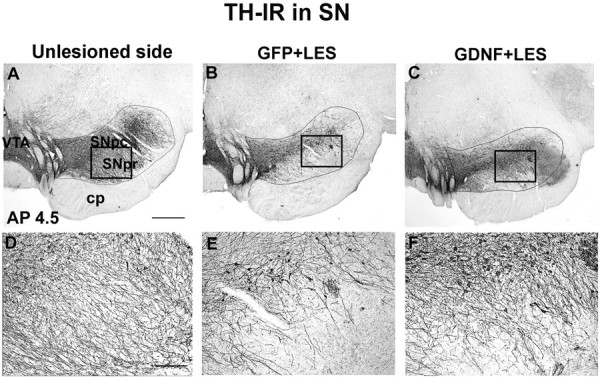
TH-IR staining of the SN (AP 4.5). A, Unlesioned SN. B, Lesioned SN in a monkey from group GFP plus LES showing substantial reduction in staining. C, Lesioned SN in a monkey from group GDNF plus LES showing partial protection against the 6-OHDA lesion. D-F, High-power micrographs taken from A-C, respectively, at the locations indicted by the boxes. E shows reduction in TH-IR fibers. F shows substantial protection of the TH-IR fibers in the SN that extend into the SN pars reticulata. SNpc, SN pars compacta; SNpr, SN pars reticulata; VTA, ventral tegmental area; cp, cerebral peduncles. Scale bars: (in A) A-C, 1 mm; (in D) D-F, 0.1 mm.