
Figure 1.
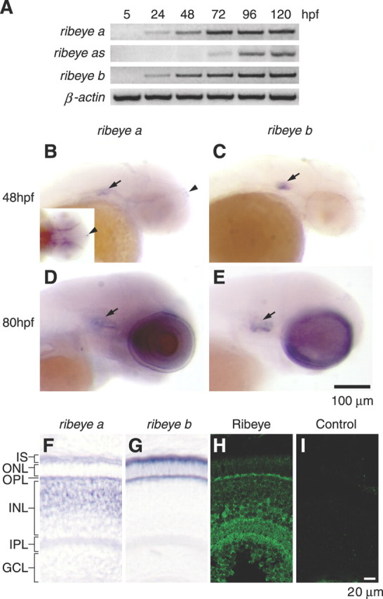
Expression of the two zebrafish ribeye genes during early development. A, RT-PCR analysis. The expression of ribeye a and ribeye b was detected at 24 hpf, whereas ribeye a2 expression was not detected until 72 hpf. β-Actin was used as a control. B, D, Whole-mount in situ hybridization analysis of ribeye a at 48 hpf (B) and 80 hpf (D). The transcripts of ribeye a were found in the inner ears (arrow), pineal gland (arrowhead and inset), and retina. C, E, Whole-mount in situ hybridization analysis of ribeye b at 48 hpf (C) and 80 hpf (E). ribeye b mRNA was identified in the inner ears (arrow) and the retina but not the pineal gland. F, G, Ribeye mRNA distribution by in situ hybridization in transverse eye sections (80 hpf). Ribeye a mRNA was found in the outer half of the inner nuclear layer, presumably bipolar cells, and in the outer nuclear layer in photoreceptor cells (F). Ribeye b was found in photoreceptor cells only (G). H, I, Immunofluorescence labeling of CtBP and Ribeye in transverse eye sections. Ribeye proteins were preferentially localized in the outer and inner plexiform layers, likely in the presynaptic terminals of photoreceptors and bipolar cells. Retinal layers are indicated as follows: IS, inner segment; ONL, outer nuclear layer; INL, inner nuclear layer; GCL, ganglion cell layer.