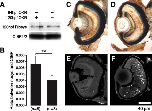
Figure 9.
Recovery of Ribeye a expression restores retinal defects. A, Western blot analysis of Ribeye a levels in recovered embryos and in OKR-negative embryos. B, Bargraph indicating that significantly more Ribeye a is present in the recovered animals (**p < 0.01; ttest) (B). C,D, Immunohistological graphs indicating that recovered embryos also have more PKCα-positive bipolar cells (C) than OKR-negative embryos (D). E, F, Fluorescence micrographs showing that apoptosis is limited in retinas of recovered embryos (E) but extensive in retinas of nonrecovered animals (F).

