Figure 3.
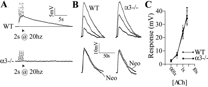
The absence of slow EPSPs but normal muscarinic responses in α3–/– ganglia. A, Stimulating the preganglionic nerve with a 20 Hz train for 2 s in the presence of hexamethonium (100 μm) produced a slow depolarization in a P7 WT neuron (top). A similar 20 Hz train stimulation produced no detectable change in membrane potential in a P7 α3–/– neuron (bottom). The stimulus artifacts were removed for clarity. Experiments were done at 37°C. B, Depolarization of a P7 WT SCG neuron (left) and a P8α3–/– SCG neuron (right) induced by three ACh concentrations (0.1×, 1×, and 5×) applied directly to each ganglia. [1× represents the concentration of ACh that produced a similar depolarization to that after preganglionic nerve stimulation (2 s at 20 Hz in neostigmine) and was usually 100μm added to the perfusion solution.] Below shows response to 1× ACh with and without neostigmine (Neo). C, ACh dose–response relationship for 11 WT (•) and 9 α3–/– (▪) neurons. The values are the means ± SEM; n = 11 for WT and 9 for α3–/–. There is no significant difference in the muscarinic response between neurons in WT and α3–/– ganglia. All experiments were done in the presence of hexamethonium (100 μm) and at 37°C.

