
Figure 5.
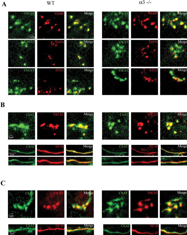
The preganglionic varicosities in α3–/– ganglia contain ChAT, VAChT, and SNARE (soluble N-ethylmaleimide-sensitive factor attachment protein receptor) proteins and are similar to those in WT ganglia. Immunostaining of varicosities in WT ganglia (left set) and α3–/– ganglia (right set) for VAChT, synaptobrevin (VAMP), syntaxin 1a (Syn1a), synaptotagmin (STG1), and ChAT. In A, the left columns show immunostaining for VAChT (green), the middle columns show immunostaining for VAMP (synaptobrevin) (top, red), syntaxin 1a (middle, red), and synaptotagmin (bottom, red), and the right columns are the merged images of the left and middle columns. The top three sets of panels show that VAMP, syntaxin 1a, and synaptotagmin colocalize with VAChT in presynaptic varicosities in P7WT and α3–/– ganglia. Scale bar, 1 μm. In B, the left columns show immunostaining for ChAT (green), the middle columns show immunostaining for VAChT (red) in presynaptic varicosities (top panels) and neurofilament (Nf-H, red) along preganglionic axons (bottom 2 panels). The right columns are the merged images of the left and middle columns and show that ChAT is present in preganglionic axons and in presynaptic varicosities in P7 WT and α3–/– ganglia. C, Similar to B but from P21 WT and α3–/– ganglia. Scale bars (in B, C): 1 μm for the varicosities; 3 μm for axons.