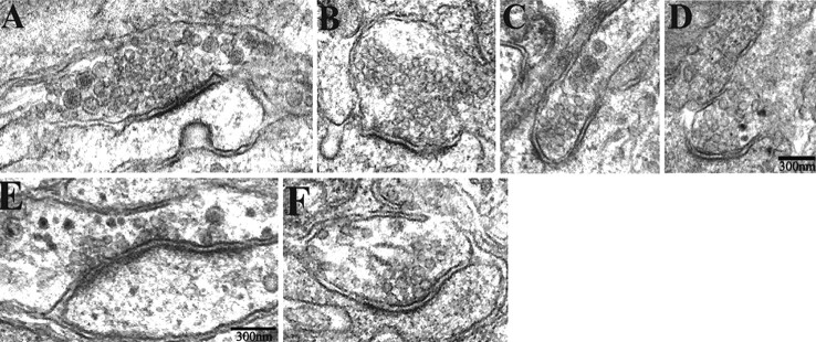
Figure 6.
The ultrastructure of synapses in α3–/– ganglia are similar to those in WT ganglia. A–F, Electron micrographs of synapses in P7 in α3–/– (A, C, E) and WT (B, D, F) SCG. The synapses have the characteristic morphological features: accumulations of synaptic vesicles adjacent to the presynaptic membrane, enhanced postsynaptic density, a parallel arrangement and thickening of the presynaptic and postsynaptic membranes, and a widened synaptic cleft. Scale bars, 300 nm.

